Why Imaging Quality Determines Surgical Outcome
A patient-specific implant is only as accurate as the imaging data on which it is based. In maxillofacial reconstruction, inadequate CT data reduces geometric detail or limit refinement and creates predictable intraoperative failure as well. When implant geometry is inaccurate, implants may rock on bone or leave gaps that compromise primary healing. When cortical boundaries and marrow space are poorly resolved, fixation becomes unreliable and screws may enter voids, tooth roots, or thin bone with poor purchase. For this reason, CT data functions as the geometric ground truth for segmentation, reconstruction, fixation planning, and final dimensional verification. Intraoperative fit is ultimately judged against this data, not against the design file.
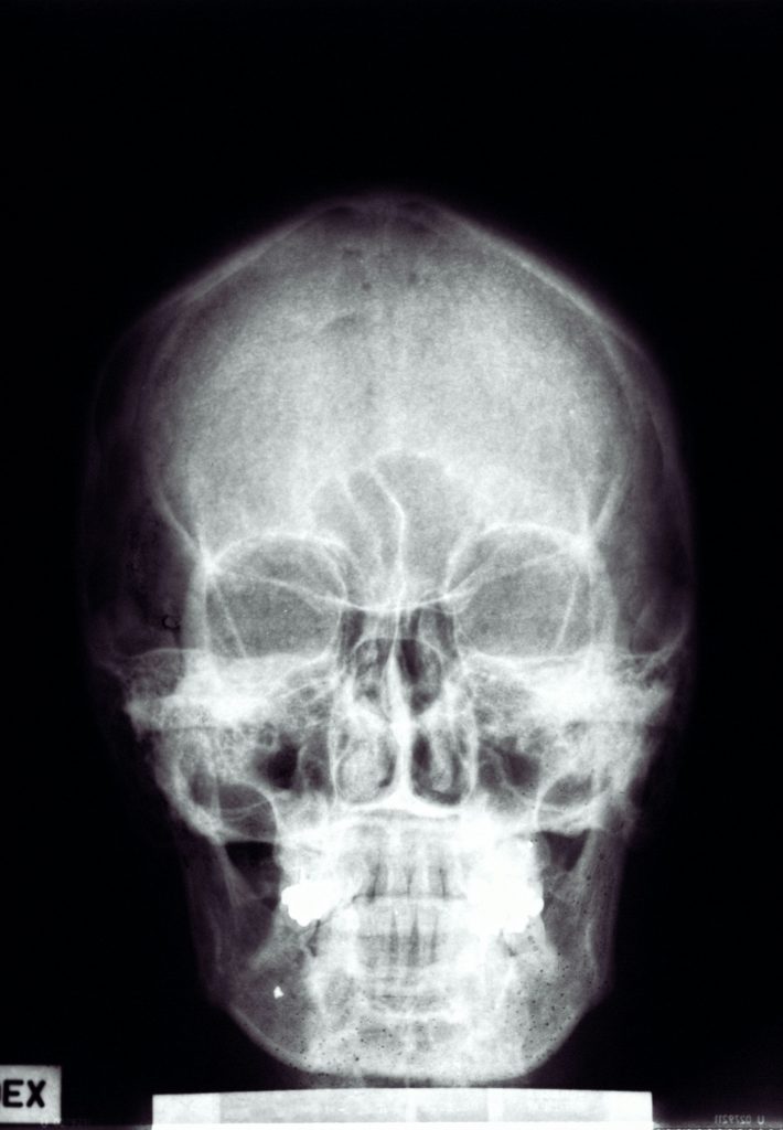
CT as the Geometric Ground Truth
In patient-specific implant workflows, CT imaging is not a reference illustration. It is the authoritative geometric representation of the patient anatomy from which all downstream decisions are derived. Errors or approximations introduced at this stage propagate directly into implant geometry and fixation strategy. Slice thickness greater than 1.0 mm introduces stair-stepping artefacts that make accurate reconstruction of fine anatomical structures such as the orbital floor, mandibular condyle, or fixation corridors unreliable, regardless of software tools or post-processing. These artefacts are a common root cause of implants that fail to seat as intended.
Recommended CT Acquisition Parameters
To minimise intraoperative surprises, CT acquisition should meet a clear technical baseline. Imaging data should be provided in raw DICOM format using a bone-optimised reconstruction kernel. Slice thickness of 1.0 mm or less is required, with 0.75 mm preferred and effectively mandatory for fixation-critical cases such as mandibular, TMJ, and orbital reconstructions. The field of view should extend beyond the immediate defect to capture relevant anatomical landmarks, ideally including the vertex, chin, and bilateral reference points. This allows stable definition of midline orientation and occlusal alignment and reduces downstream uncertainty during reconstruction.
Common Data Limitations and Their Consequences
When imaging falls short of these conditions, the resulting consequences are well understood. Metal artefact from existing plates, screws, or dental restorations can obscure cortical margins and screw trajectories, increasing the risk of fixation failure unless artefact-reduction techniques or manual segmentation are applied with explicit surgeon confirmation of boundaries. Thick-slice imaging reduces conformity at the bone-implant interface and often necessitates deliberately looser design tolerances, with acceptance of increased intraoperative contouring. Partial field-of-view acquisition limits mirroring and symmetry assessment and frequently leads to late design revision or increased intraoperative adjustment, particularly when reference anatomy is missing outside the defect region.
MRI and Multimodal Imaging
MRI is not used as a primary dataset for implant geometry. However, MRI may be co-registered with CT where soft tissue extent, tumour margins, or flap bulk are expected to influence implant contour, clearance, or fixation access. In such cases, MRI informs design constraints rather than defining implant geometry directly. All dimensional and fixation decisions remain grounded in CT data.
Clinical Reality and Decision-Making Under Constraint
In trauma and oncological cases, ideal imaging is not always achievable. Delaying surgery for repeat acquisition may be clinically unacceptable. In these situations, proceeding is a deliberate decision rather than a default response to suboptimal data. When imaging limitations remain, design strategy is adapted to prioritise predictable fixation and intraoperative behaviour over geometric refinement or aesthetic optimisation. No design progresses to manufacture without an initial data review. Where imaging constraints persist, the resulting design assumptions are discussed directly with the surgeon and accepted knowingly, with fixation strategy adjusted accordingly.
Closing Perspective
Patient-specific implant design is not a process of fixing inadequate data with advanced software. It is a process of preserving accuracy where possible and making deliberate, shared compromises where clinical reality requires them. Predictable surgical outcomes begin in the radiology suite, not in the operating room.